
The Imaging and Characterization Core Lab houses fifteen electron microscopes. Several of these microscopes enable sub-angstrom imaging capability and ultra high-energy resolution chemical analysis capability.
Recently, the lab creatively applied a technique usually used for biological samples to solve challenging material science issues. This enabled us to capture the first-ever atomic-resolution TEM image of an ultra-beam sensitive material (MOF). This resulted in the publication of “Unravelling surface and interfacial structures of a metal-organic framework by transmission electron microscopy ” in Nature Materials. Core Labs staff worked with KAUST’s Advanced Membranes and Porous Materials Center, collaborators from Gatan, the Lawrence Berkeley National Laboratory and Xi’an University of Science and Technology. Learn more about this breakthrough via KAUST Discovery.
Core Labs scientists are developing novel imaging techniques that will make imaging beam-sensitive materials routine in the near future.
We also recently used our Transmission Electron Microscopes (TEM) to create a new way to conduct DPC imaging. For decades, researchers have been performing differential phase contrast (DPC) imaging using dedicated equipment and software. Recently, staff from the Imaging and Characterization Core Lab have identified a way to conduct DPC with standard equipment only, making the DPC method widely available. Learn more about this new methodology which is already getting attention in research groups and has resulted in several publications.
Planning for the future
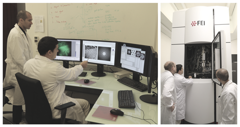
FEI’s Titan Themis TEM is a double-corrected instrument that provides atomic-scale imaging and analysis. It will be used by materials scientists to understand relationships between a material’s larger-scale physical properties and its atomic-scale composition and structure. With future discoveries in mind, the Imaging and Characterization Core Lab will soon be home to a new industry-leading TEM. The Titan Themis Z will enable scientists to view and identify single atoms according to their atomic number. Scientists will be able to use this new instrument to characterize materials at the atomic scale and study their catalytic, magnetic and electrical properties.
Get started
The Imaging and Characterization Core Lab specializes in spectroscopy and microscopy for material and device research to learn about the structure and composition of samples. It includes facilities for electron microscopy, nuclear magnetic resonance, physical characterization, surface science, and optical microscopy. The facility enables researchers to view samples at a scale ranging from millimeter to sub-angstrom, providing a wide spectrum of imaging and characterization capabilities.
The Titan Themis Z will be operational this spring and will be supported by scientists and specialists from our electron microscopy facility.
Our services are available for use by the KAUST research community, collaborators and industrial partners. For more information, contact us.