
Delivering a wide spectrum of capabilities in imaging, structure, and spectroscopy characterization
The Imaging and Characterization Core Lab houses state-of-the-art technologies in the disciplines of electron microscopy, nuclear magnetic resonance, physical characterization, surface science, and optical microscopy.
Watch Video
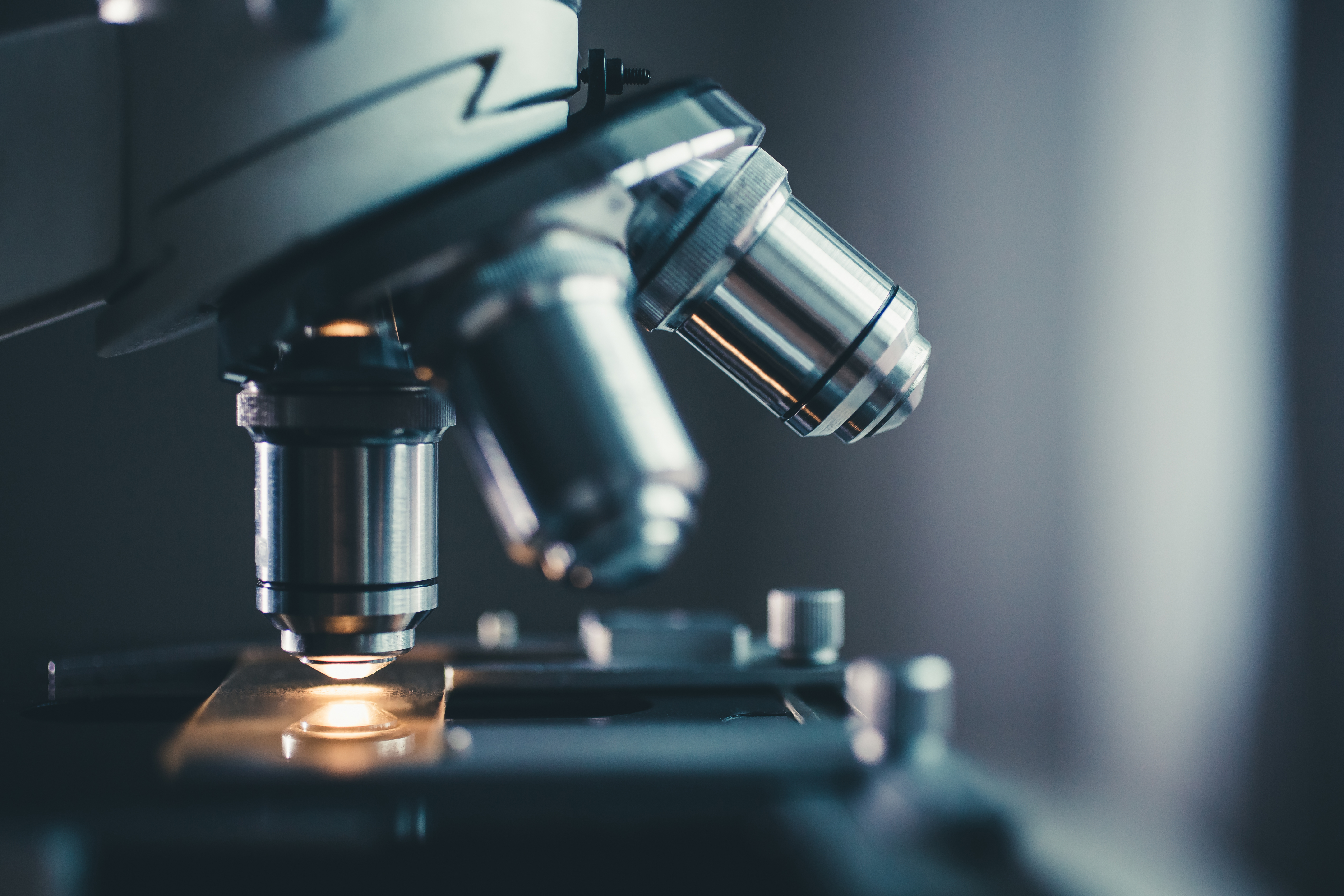
The optical microscopy lab houses advanced optical microscopes which provide capabilities in both biological and materials science imaging and characterization. Our expert staff specialize in: cell culturing; staining and tissue processing; 2D, 3D and 4D imaging; advanced live cell imaging; image processing; surface topography inspection; grain size and porosity analysis; and dynamic process imaging and physical characterization.
Within this area a Center of Excellence for Optical Microscopy has been established between KAUST and Leica Microsystems. This partnership supports cutting-edge life science research.

For a listing of technologies in this area please click here
Our NMR lab, supported by our partnership with Bruker, presents cutting edge advanced technologies for obtaining atomic resolution information on the structure and motion of molecules. Processes we are proficient in include; one-dimensional, multi-dimensional, and dynamic nuclear polarization NMR spectroscopy, electron paramagnetic resonance spectroscopy and magnetic resonance imaging for macroscopic images.

For a listing of technologies in this area please click here
The physical characterization lab specializes in physical property characterization, with a focus on X-ray diffraction, raman spectroscopy, and electrical characterization. Our expertise includes: phase identification of polycrystalline samples by powder X-ray diffraction, 3-D structure determination of single crystals, structural properties measurement of epitaxial thin film, Raman molecular group identification, photoluminescence (PL) analysis for light emission position determination and electrical probing of wafers, dice, and packaged devices.

For a listing of technologies in this area please click here
Our facility specializes in a comprehensive suite of advanced electron microscopy techniques for solid and stable materials, as well as soft and beam sensitive. Thenabling high-resolution imaging, in-depth chemical analysis, and structural characterization at the atomic scale. Our expertise includes:
•3D imaging via conventional FIB, Plasma FIB, and Cryo-FIB.
With these state-of-the-art capabilities, we empower researchers across disciplines to push the boundaries of materials science, nanotechnology, and structural biology
For a listing of technologies in this area please click here
Surface science deals with the property of surfaces and sub-surfaces up to a depth of several nanometers. This lab is equipped with technologies for analyzing surface morphologies, chemical states, chemical compositions and composition distribution along depth, enabled by X-ray photoelectron spectroscopy, secondary ion mass spectrometry (SIMS) and atomic force microscopy.

For a listing of technologies in this area please click here