
---khulud-m-_chu6460102-resized.tmb-th950x345.jpg?Culture=en&sfvrsn=fd7a413b_1)
By the Imaging and Characterization Core Lab team and Caitlin Clark
In March 2018, KAUST and Leica Microsystems established a Center of Excellence (CoE) for Optical Microscopy on the University's campus. The CoE is housed in the Imaging and Characterization Core Lab (IAC).
“The CoE places KAUST at the leading edge of technological advances in optical microscopy—an auspicious partnership that will positively impact the entire KAUST academic community, its collaborators and industrial partners by encouraging new discoveries in life science research,” stated Dr. Daniel Acevedo-Feliz, facilities director of Materials Characterization, Nanofabrication and Plant Science Core Labs.
Planning strategic objectives
As part of the implementation of the CoE, a KAUST-Leica strategic board meeting took place on campus in March 2019. During the meeting, Leica introduced its most advanced microscopy technology and the company’s future technical roadmap. Participants also discussed streamlining Leica’s technology to KAUST’s research requirements.
A series of actions, including setting up remote STimulated Emission Depletion (STED) microsystem diagnosis and four level maintenance, was put into place to further improve maintenance service for the KAUST research community.
In the future, the KAUST-Leica CoE plans to organize periodic educational workshops and high-level technical forums. These will improve the mutual technical understanding between KAUST researchers and Leica Microsystems, which will lead to tailor-made optical microscopy technology for KAUST research purposes and collaborative optical imaging methodology development.
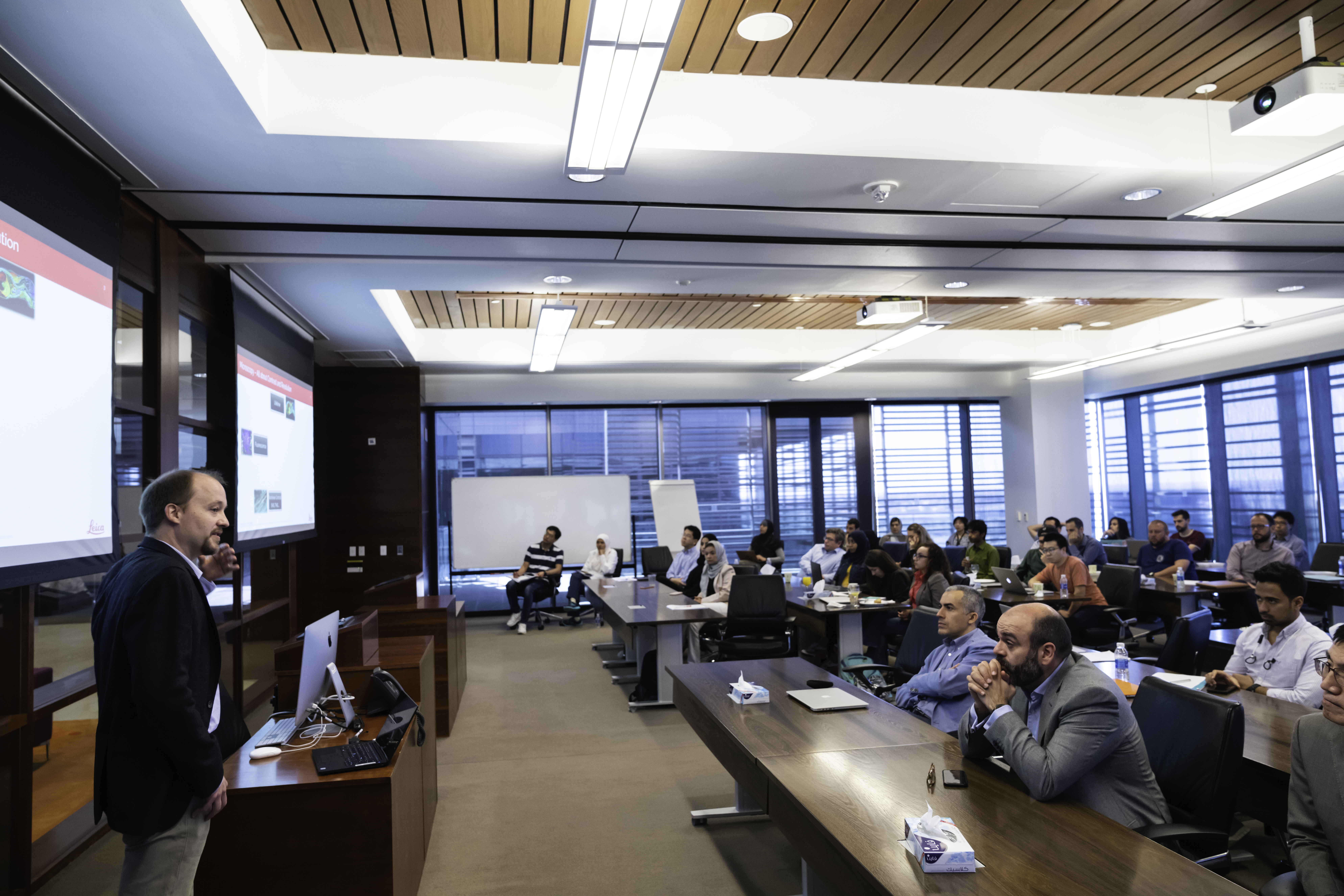
Dr. Volker Schweikhard, product application manager EMEA, Leica Microsystems, gives a presentation to attendees of a KAUST Imaging and Characterization Core Lab-Leica biological technical forum in March 2019. File photo.
The evolution of cell understanding—advanced biological optical microscopy technical forum
Along with the strategic board meeting, the IAC and Leica Microsystems cohosted a biological technical forum. The forum focused on introducing the newest optical microscopy commercial advances in confocal microscopy, deep tissue high resolution imaging, label-free live cell imaging, fluorescence life time imaging and haze free wide field imaging techniques. It also introduced KAUST’s biological research progress in super-resolution microscopy, live cell imaging and plant science studies.
The forum was attended by over 50 KAUST scientists, including students, postdoctoral fellows and faculty members, and it also included several Leica application experts, who discussed the most recent Leica microscopy technologies.
During the event, KAUST Professors Carlo Liberale, Satoshi Habuchi and Ikram Blilou also shared their newest research results in Raman scattering, microfluidic-based super-resolution and multiscale plant tissue study. Attendees also had the opportunity to test their samples by using Leica super-resolution STED and the digital light sheet microscope installed in the IAC.
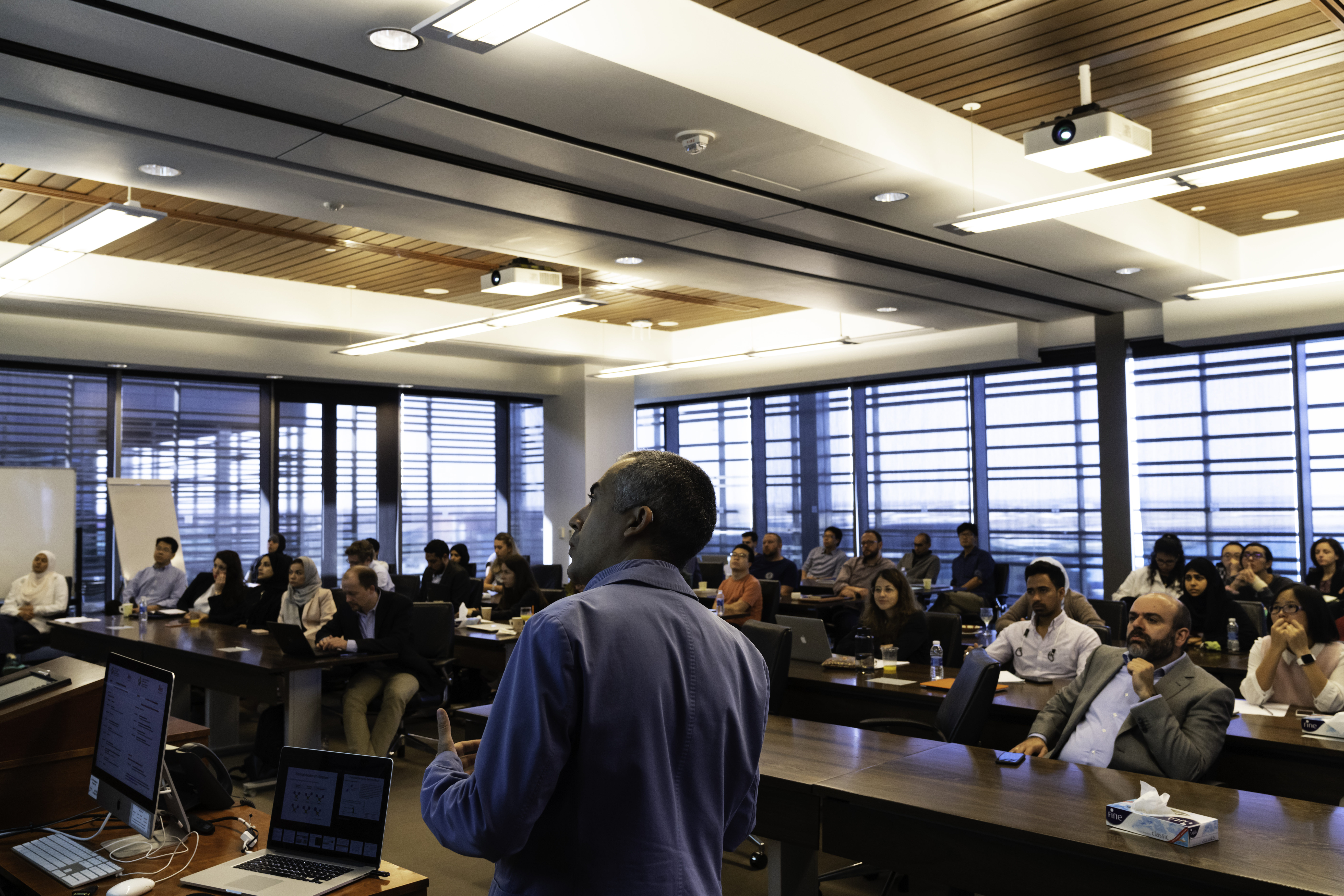
Dr. Carlo Liberale, KAUST assistant professor of bioscience, presents on broadband stimulated Raman scattering microscopy during a KAUST Imaging and Characterization Core Lab-Leica biological technical forum in March 2019. File photo.
Product demos for researchers
In May 2019, the KAUST-Leica CoE held a two-week hands-on testing product demo for LIGHTNING and PAULA to promote advanced biological optical microscopy techniques.
The Personal AUtomated Lab Assistant (PAULA) helps to speed up daily cell culture tasks and standardize the results to improve downstream workflows, offering essential analysis apps like confluence efficiency check. It also helps to ensure that cultivated cells are ready for the next step and that everything is well-documented.
LIGHTNING helps to maximize the information extracted from a specimen and get in-depth answers to scientific questions. Through the use of LIGHTNING and with one click, it can reveal the most information from the images taken and resolve nanoscale structures down to 120 nanometers.
More than 15 KAUST researchers attended the hands-on demo, and they were deeply impressed by the new techniques. They learned about PAULA’s ease of use, fast analysis and capability to free up valuable work time for scientists. The researchers felt that PAULA is the best technique to replace the manual and time-consuming traditional cell culture analysis microscope used in the lab.
Researchers also experienced the image quality improvement brought by LIGHTNING technology.
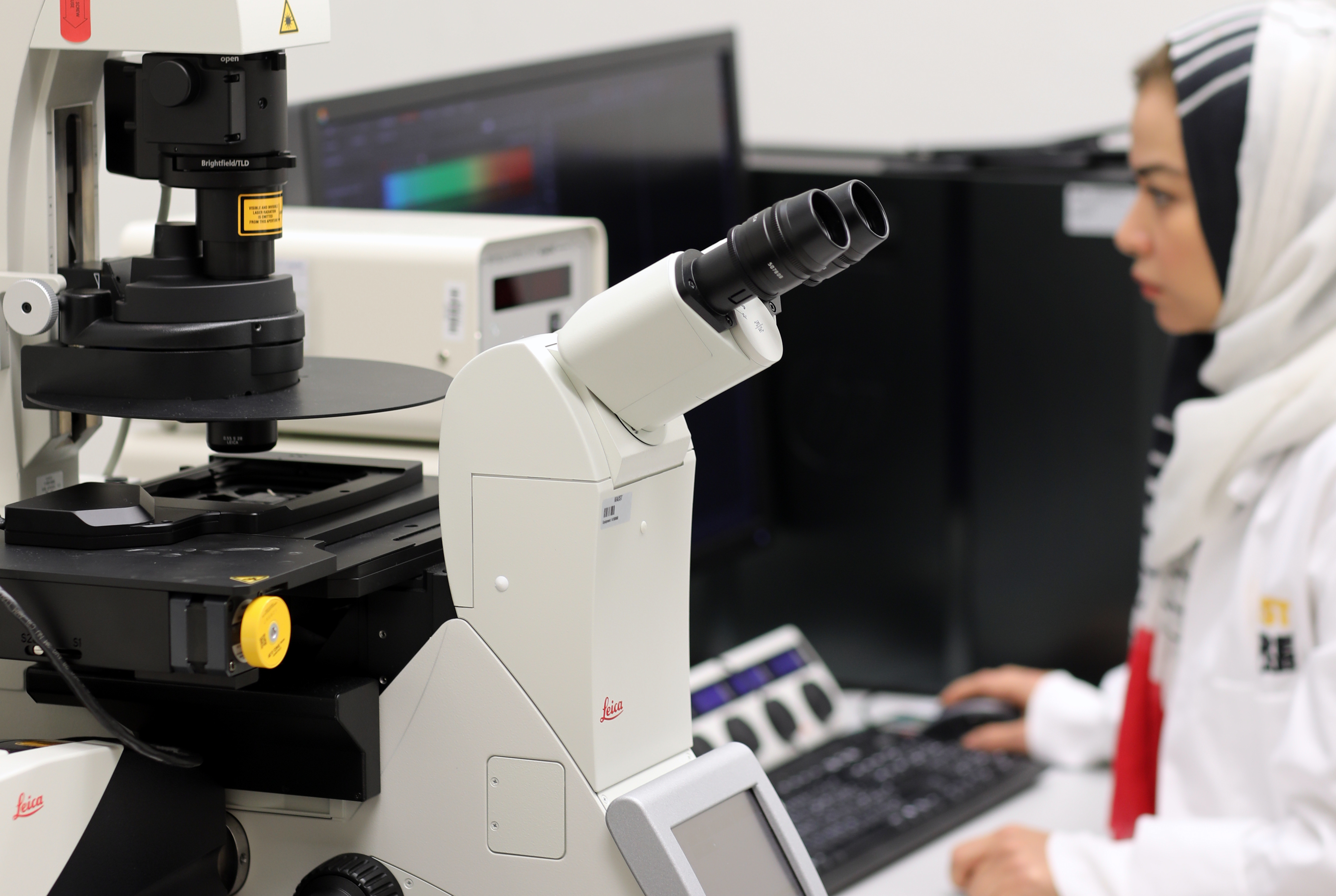
A KAUST researcher makes use of technology from Leica Microsystems on campus. File photo.
Xuan Zhou, a KAUST biological science Ph.D. student from University Professor Mo Li’s research group, obtained a stunningly clear cellular image using LIGHTNING; the image featured many more cell structure details compared to blurry images previously obtained using a traditional confocal microscope.
"My lab is interested in studying the pathological changes in the nucleolus, a tiny subnuclear organelle a few microns in diameter, in immune cells of patients with Wiskott-Aldrich syndrome, a devastating immunological disease,” explained Li, who is an assistant professor of bioscience.
“Traditionally, the compartments of the nucleolus can only be revealed by electron microscopy,” he continued. “Our efforts to visualize the compartments using conventional confocal microscopy failed to provide satisfactory results. My students received training from the Core Labs staff and Leica specialists on the Leica STED 3X and successfully imaged all three compartments with excellent resolution. The LIGHTNING capability further enhances the image quality of STED and is a breeze to work with. I hope these images can give our two upcoming manuscripts an edge in the publication process.”
"Leica’s LIGHTNING technology offers fast and high-quality images,” noted Zhou. “The STED plus LIGHTNING technology gave us the essential details inside the nucleolus that we never observed before. Without a doubt, LIGHTNING is a very powerful tool for our research—we can see the structure and we know what we see is true."
"Leica LIGHTNING technology is fantastic,” added Baolei Yuan, a KAUST Ph.D. student in biological science who is also from Li’s group. “It helped me harvest high-resolution images with more details of protein locations and structures, which are hard to observe using traditional technologies. The white light laser and hybrid detectors of Leica SP8 are also very helpful for me to make staining easier without worrying too much about the dye combinations and weakness of signal."
The product demo successfully helped KAUST researchers to learn the newest microscopy technology and provided more alternatives for the devices and techniques to implement for their future research work.
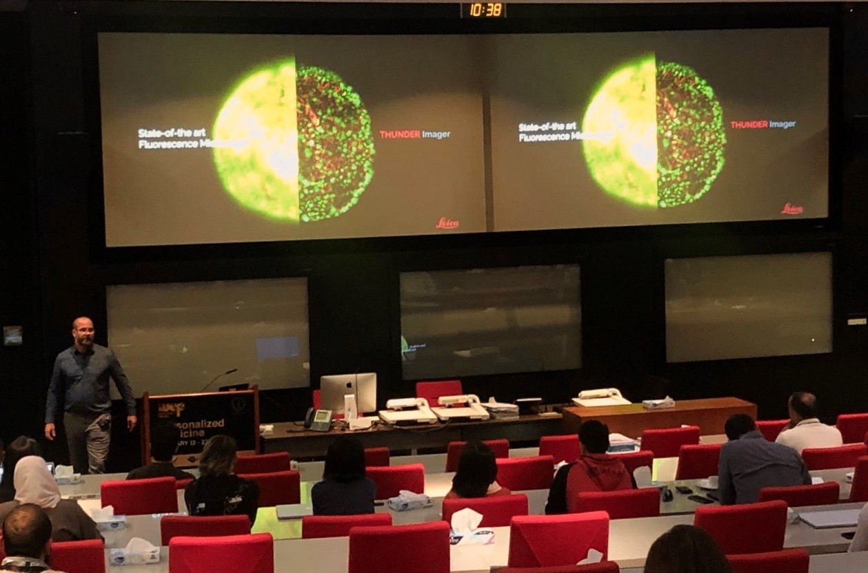
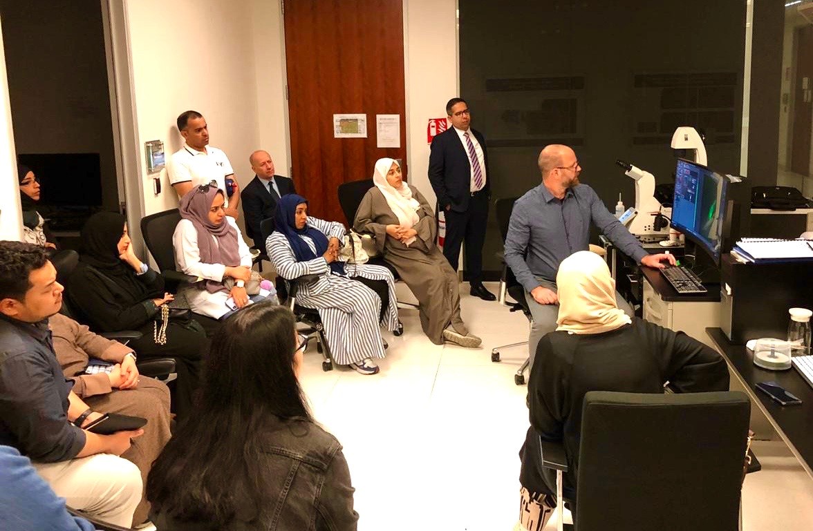
Dr. Jan Schumacher, advanced workflow specialist, Leica Microsystems Germany, showcases THUNDER technology during the Advanced Fluorescent Microscopy workshop held at KAUST in January 2020. File photo.
Advanced Fluorescent Microscopy Training Course
In January 2020, KAUST and Leica hosted a two-day workshop/training course on the University’s campus that focused on advanced optical microscopy technologies and how these can be applied in life science research to enhance and optimize workflows for image acquisition.
The course consisted of technical presentations and hands-on sessions, during which users had the opportunity to try out their own samples on the THUNDER Demo system and on the KAUST SP8X STED system currently available in the IAC.
After completing the course, a total of 45 scientists and researchers from KAUST and other Saudi research institutions, including KAU/KFMRC, IMC Hospital, King Khalid University and Jazan University, obtained a solid understanding of cutting-edge techniques in high-end fluorescence and confocal microscopy.
Based on participants’ comments, the workshop was extremely helpful in providing valuable insight and hands-on time on advanced equipment. The IAC also conducted a post-workshop satisfaction survey for KAUST users, which resulted in many positive comments. The users were pleased with having LIGHTNING technology available in the Core Lab, and the event triggered a high interest in having the newest Tau-STED and THUNDER technology showcased to improve image acquisition and analysis in ongoing research projects.
A hands-on and product demonstration of THUNDER technology led by Dr. Jan Schumacher (right, seated), advanced workflow specialist, Leica Microsystems Germany, took place on campus in January 2020. File photo.
New horizons for the KAUST-Leica CoE
Leica’s technology has proven to be beneficial for life science research on campus. The plan for the future is to utilize KAUST as a point of reference in the Kingdom for Leica’s latest innovative technologies and to inspire new collaborations with in-Kingdom research institutions and industry partners.
“We as Leica Microsystems are committed to supporting KAUST in every dimension—not only in daily research activities but also through the initiatives that KAUST is and will be leading in accordance with Saudi Vision 2030,” stated Tunç Koşcağız, Leica Microsystems’ sales director for the Middle East, Africa and South East Europe.