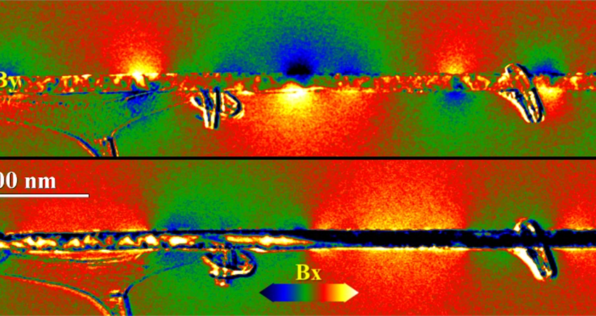
For decades, researchers have been performing differential phase contrast (DPC) imaging using dedicated equipment and software. Recently, staff from the Imaging and Characterization Core Lab have identified a way to conduct DPC with standard equipment only, making the DPC method widely available.
DPC is a method in Transmission Electron Microscopy (TEM) that visualizes and quantifies local magnetic and electrostatic fields in a range of nanomaterials. The method is useful for fast switching between studying objects of about 10µm down to a few nm in size. Core Lab staff collaborated on the development of the Virtual Bright Field DPC (VBF-DPC) method by combining a new concept of unitary DPC detector developed by Sergei Lopatin and automated data acquisition and processing developed by Daliang Zhang.
The implementation of the new VBF-DPC method is already getting attention in research groups and has resulted in several publications, including:
-
Ivanov, Yurii P; Chuvilin, Andrey; Lopatin, Sergei; Kosel, Jurgen; Modulated Magnetic Nanowires for Controlling Domain Wall Motion: Towards 3D Magnetic Memories, ACS nano, 10 (5), 5326–5332, 2016
-
Lopatin, Sergei; Ivanov, Yurii P; Kosel, Jurgen; Chuvilin, Andrey; Periodic Magnetization Pattern for Controlled Domain Wall Motion in Nanowires, Microscopy and Microanalysis, 22 (S3), 1678-1679, 2016
-
Lopatin, Sergei; Ivanov, Yurii P; Kosel, Jurgen; Chuvilin, Andrey; Multiscale differential phase contrast analysis with a unitary detector, Ultramicroscopy, 162, 74-81, 2016
The video above was captured using the new VBF-DPC method. It shows the 3D visualization (tomography) of magnetic domains in a nanowire made of Ni and Co segments. The visualization is performed in a temperature color scheme. The dark colors (blue-black) correspond to the magnetic field pointing down and to the left. The darker the color, the stronger the magnetic field is. Bright colors (yellow-white) correspond to the magnetic field pointing upward and to the right.

