
By the Imaging and Characterization Core Lab team and Marian O'Neill
They say that beauty is in the eye of the beholder. In the art of sample preparation this maxim holds a special truth. As every scientist knows a correctly prepared sample presents the beauty of discovery, of understanding, of wonder to the beholder; three essentials to research.
From Tuesday Nov 14th to Thurs Nov 16th the KAUST Imaging and Characterization Core Lab hosted from Eye to Insight. This workshop was the first intensive workshop held in the Middle East to present the forefront of microscopy technology in tandem with demonstrations in sample preparation. It was presented in collaboration with Leica Microsystems and their distributor Salehiya Establishment. Thirty four students attended: as well as faculty members and researchers from KAUST, attendees travelled from Umm Al-Qura University, University of Jeddah, King Abdul Aziz University and Express Lab Bahrain.
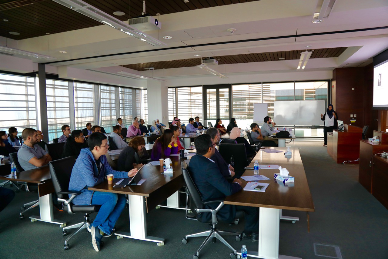
Ptissam Bergam from KAUST lecturing at From Eye to Insight. Photo taken by Rachid Sougrat
With cryo TEM being awarded the Nobel Prize in chemistry in 2017 for the structural determination of bio molecules, this workshop has proved timely. TEM, SEM and cryo technologies are integral to opening our eyes to the subtleties and secrets held at atomic level. But, no matter how high a resolution these technologies are capable of attaining, results are always dependent on the quality of the sample.
Designed to be as informative, interactive and engaging as possible From Eye to Insight was structured to deliver theory in the morning and related, hands on demonstrations in the afternoon. Both lectures and demonstrations covered the full spectrum of microscopy, from the biological to the material sciences; teaching students, from diverse backgrounds, how to prepare samples that result in great, rather than good images.
Dr Sougrat, from the KAUST Imaging and Characterization Core Lab, delivered the opening lecture; an exploration into the art of sample preparation in electron microscopy. A skill heightened to the definition of art because of the informed level of dexterity it demands.
Other lectures on the morning of the first day, delivered by Dr Nowak from Leica and Ptissam Bergam from KAUST, covered; room temperature sample preparation techniques – ultra thin sectioning for TEM applications and cryo Ultramicryomy/ Tokuyasu Technique & Immunogold Labelling for TEM applications.
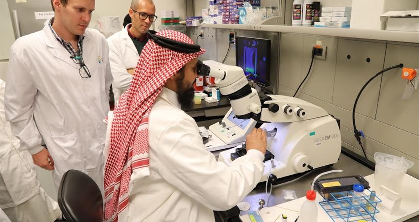
After a tour of the Imaging & Characterization Core Lab the first series of afternoon demonstrations explored room temperature ultramicrotome procedures and cryo sectioning.

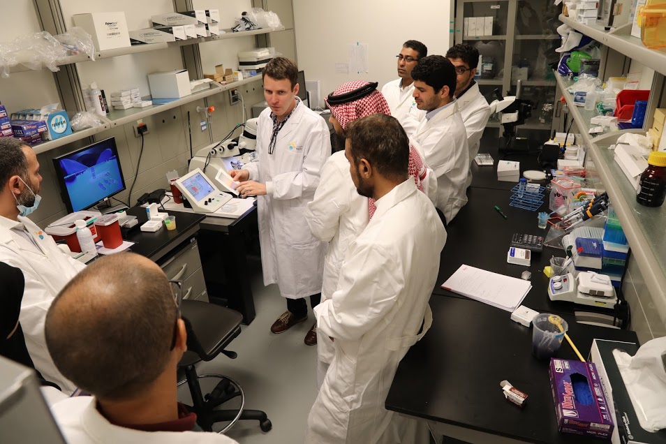
Hands on demonstrations in the KAUST Imaging and Characterization Core Lab. Photo taken by Rachid Sougrat
Dr Nowak opened the lectures on day two with an investigation into cryo SEM sample preparation workflow. His colleague from Leica, Dr Leroux, followed with a talk on coating techniques for SEM applications including the REPLICA technique. Dr Behzad from KAUST closed this lecture session with his talk on cryo SEM principles and applications.
As the morning lectures indicated the afternoon demonstrations were all about the SEM. Dr Behzad led with an investigation into Cryo SEM and a discussion of its applications. Dr Leroux followed demonstrating freeze fracture, high vacuum cryo coating and sample transfer to the cryo SEM.
Day three concentrated on chemical fixation and high pressure freezing. Dr Sougrat opened with a lecture on the chemistry of chemical fixation. This was followed by Dr Leroux's talk on high pressure freezing – the technique and its advantages over chemical fixation. After a coffee break Dr Nowak followed up with an exploration into the latest developments in high pressure freezing. Dr Leroux closed the lecture module of this workshop with a talk on freeze substitution, the link between a frozen sample and room temperature follow-on procedure.
The final lab demonstrations showcased freeze substitution and high pressure freezing.
From Eye to Insight was highly valued by participants and presenters alike. 'Inspiring', 'informative', 'helpful' and 'brilliant' were just some of the adjectives used to describe this eye opening experience. As we all know, until very recently our understanding was limited by what the eye could see. The complexities that structure the visible fabric are only now being brought into focus. Technologies such as electron microscopy, coupled with the sample preparation techniques covered in From Eye to Insight, are redefining our perceptions.